The nervous system allows us to transform environmental stimuli into perception, thought, and action. While the spinal nerves connect the body to the spinal cord, the cranial nerves connect the head and neck directly to the brain. In this section, we'll take a closer look at the 12 cranial nerves—their roles, pathways, and importance in daily life. For more detail on neural anatomy, check out our 3D interactive models on the site.
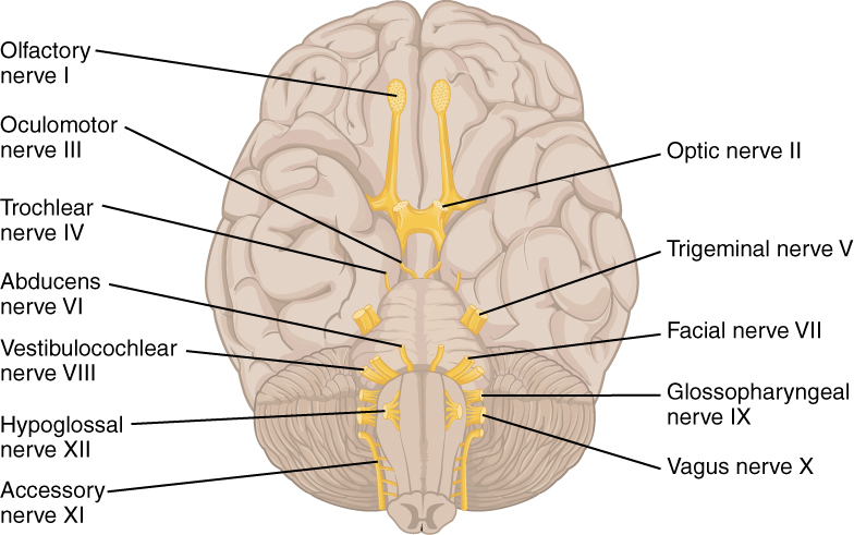
There are 12 cranial nerves, most located in the head and neck (Sonne et al., 2025). Each has specialized roles—some carry sensory information, others control muscles, and a few do both.
- Cranial Nerves I and II (Olfactory and Optic) are extensions of the central nervous system (CNS).
- The other ten (III–XII) arise from the brainstem and belong to the peripheral nervous system (PNS).
Cranial Nerve - Olfactory Nerve (I)
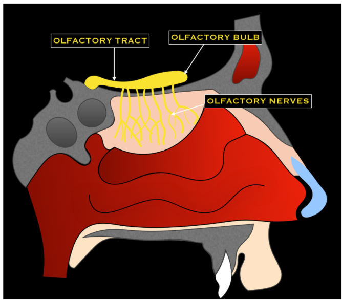
The olfactory nerve carries smell information from receptors in the nasal cavity to the brain (Romano et al., 2019). Unlike most nerves, its fibers are not myelinated by Schwann cells. Humans have about 6 million olfactory receptors, while dogs boast nearly 200 million, giving them a far superior sense of smell (Libreros-Jiménez et al., 2023). Smell is deeply tied to survival—warning us of danger (e.g., spoiled food, gas leaks)—and even plays a role in social bonding and memory (Poirier & Melin, 2023).
Cranial Nerve - Optic Nerve (II)
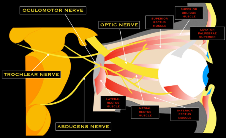
The optic nerve transmits visual information from the retina to the brain. Rods detect light and motion, while cones detect color. After phototransduction, signals pass through the optic chiasm and are processed mainly in the occipital lobe's visual cortex (Rueda-Lopes, 2021). Vision enables spatial navigation, depth perception, and environmental awareness.
Cranial Nerve - Oculomotor Nerve (III)
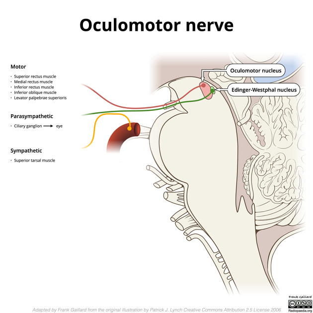
The oculomotor nerve controls most eye movements and pupil constriction. It also adjusts the lens for near and far vision. Without it, focusing and coordinated eye motion would be impossible.
Cranial Nerve - Trochlear Nerve (IV)
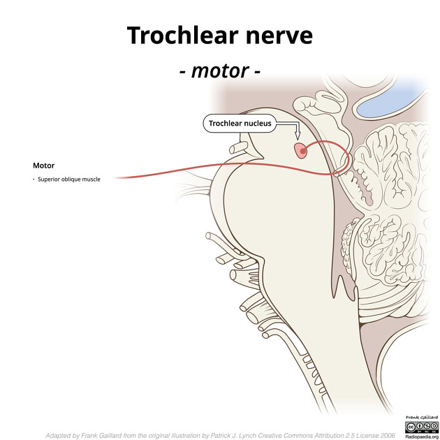
The trochlear nerve controls the superior oblique muscle, enabling downward and inward eye movement. It is the only cranial nerve that exits from the back of the brainstem. Historically, it was nicknamed the "pathetic nerve" due to its role in downward gaze (Libreros-Jiménez et al., 2023).
Cranial Nerve - Trigeminal Nerve (V)
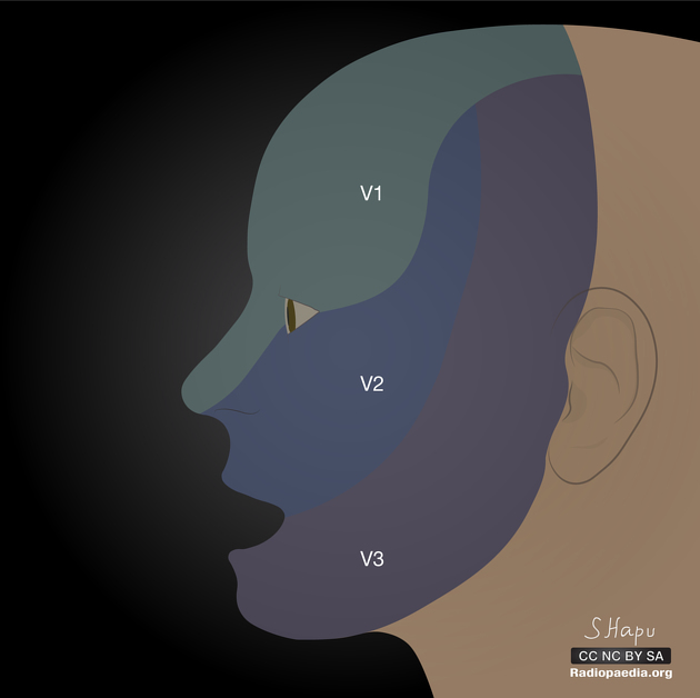
The trigeminal nerve is the largest cranial nerve, carrying both sensory and motor signals. It has three major branches:
- Ophthalmic (V1): sensation from the eyes and forehead.
- Maxillary (V2): sensation from the midface.
- Mandibular (V3): both sensory and motor functions, including chewing muscles.
Cranial Nerve - Abducens Nerve (VI)
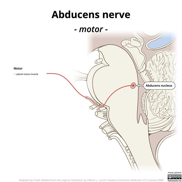
The abducens nerve controls the lateral rectus muscle, which moves the eye outward. Damage to this nerve often causes double vision.
Cranial Nerve - Facial Nerve (VII)
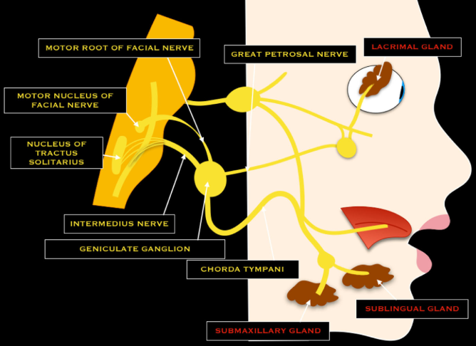
The facial nerve controls facial expression, blinking, and some neck muscles. It also carries taste sensation from the front two-thirds of the tongue and regulates tear, saliva, and mucus production. Its wide range of functions makes it vital for both survival and communication.
Cranial Nerve - Vestibulocochlear Nerve (VIII)
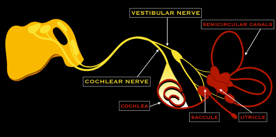
This nerve has two parts:
- Cochlear branch: transmits sound from the cochlea.
- Vestibular branch: detects head position and motion for balance.
Together, they enable hearing and equilibrium.
Cranial Nerve – Glossopharyngeal Nerve (IX)
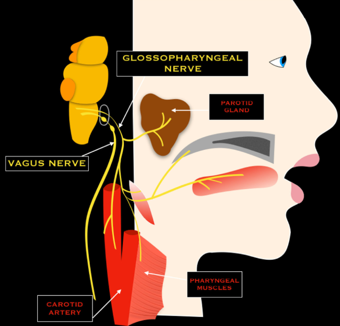
The glossopharyngeal nerve contributes to taste (posterior tongue), swallowing, saliva secretion, and sensory input from the throat and middle ear. It also plays a role in monitoring blood pressure and oxygen levels.
Cranial Nerve – Vagus Nerve (X) (Wanderer)
The vagus nerve is the body's "wanderer," extending from the brainstem down into the chest and abdomen. It regulates heart rate, digestion, breathing, and parts of the immune response. Remarkably, it supplies about 75% of parasympathetic output in the body.
Cranial Nerve – Accessory Nerve (XI)
The accessory nerve supplies motor input to neck and shoulder muscles, allowing head rotation and shoulder elevation. It has both cranial and spinal roots, working closely with the vagus nerve.
Cranial Nerve – Hypoglossal Nerve (XII)
The hypoglossal nerve controls tongue movement, essential for speech, swallowing, and chewing. It activates both intrinsic (shape) and extrinsic (movement) tongue muscles.
Functional Grouping
- Sensory only: I (Olfactory), II (Optic), VIII (Vestibulocochlear)
- Motor only: III (Oculomotor), IV (Trochlear), VI (Abducens), XI (Accessory), XII (Hypoglossal)
- Mixed (sensory + motor): V (Trigeminal), VII (Facial), IX (Glossopharyngeal), X (Vagus)
The cranial nerves are gateways between the brain and the outside world. From perceiving odors and sounds to moving our eyes, faces, and tongues, they integrate sensation, movement, and autonomic functions that sustain life. Understanding them is not only key to anatomy—it's essential for appreciating how our nervous system shapes daily experience.
References
- Libreros-Jiménez, H. M., Manzo, J., Rojas-Durán, F., Aranda-Abreu, G. E., García-Hernández, L. I., Coria-Ávila, G. A., ... & Hernández-Aguilar, M. E. (2023). On the cranial nerves. NeuroSci, 5(1), 8-38.
- Poirier, A. C., & Melin, A. D. (2024). Smell throughout the life course. Evolutionary Anthropology: Issues, News, and Reviews, 33(4), e22030.
- Romano, N., Federici, M., & Castaldi, A. (2019). Imaging of cranial nerves: a pictorial overview. Insights into imaging, 10(1), 33.
- Rueda-Lopes, F. (2021). The cranial nerves: extensions of the central nervous system or components of the peripheral nervous system-how should we evaluate them?. Radiologia Brasileira, 54(3), V-VI.
- Sonne J, Lopez-Ojeda W. Neuroanatomy, Cranial Nerve. In: StatPearls. StatPearls Publishing, Treasure Island (FL); 2025. PMID: 29261885.