Spinal Column (Bone)
The spine consists of a series of small bones (vertebrae) that protect the spinal cord (Yu et al., 2020). It includes 24 articulated (mobile) vertebrae separated by intervertebral discs, and 9 non-articulated (fused) vertebrae found in the sacral and coccygeal regions. Details about each vertebra are provided in Table 1 within the appendices.
Spinal Nerves (Neurons)
The spinal cord is a cylindrical structure that emerges from the medulla oblongata (where the spinal cord meets the skull) and travels down the spine. Pairs of spinal nerves exit and enter through each vertebra (left and right), forming part of the peripheral nervous system (PNS) (Yu et al., 2020; Kaiser & Lugo-Pico, 2019). These spinal nerves transmit sensory information to the central nervous system (CNS) and initiate motor responses.
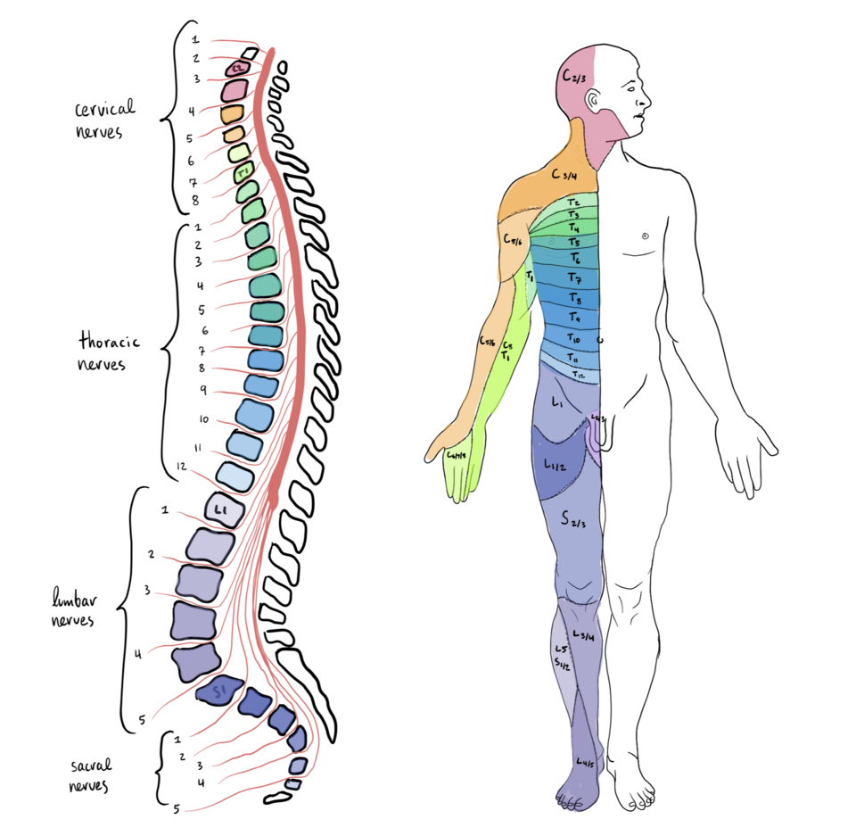
There are 31 pairs of spinal nerves connecting the CNS and PNS:
- 8 cervical pairs (C1–C8, neck region)
- 12 thoracic pairs (T1–T12, chest region)
- 5 lumbar pairs (L1–L5, lower back)
- 5 sacral pairs (S1–S5, pelvis)
- 1 coccygeal pair (Co1, tailbone)
Each spinal nerve is a mixed nerve, containing both sensory (afferent) and motor (efferent) fibers, which extend outward to form muscle fibers, receptors, and components of the autonomic nervous system (ANS).
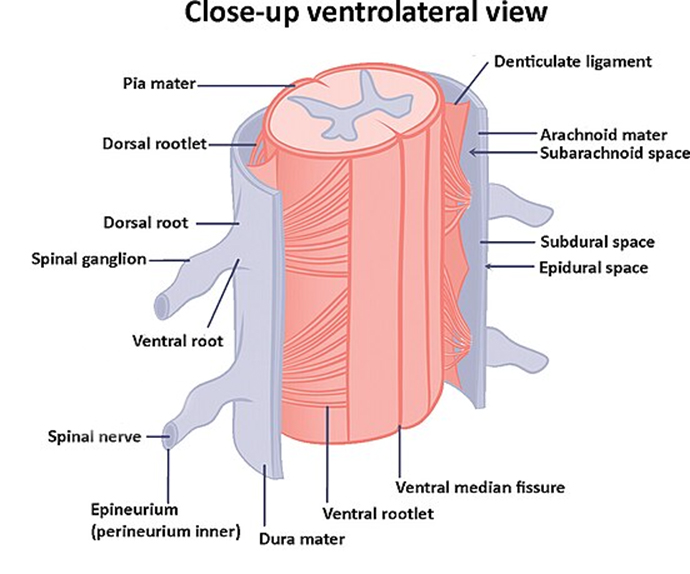
Key Structures of a Spinal Nerve
- Spinal Nerve: Formed by the union of dorsal (sensory) and ventral (motor) roots; exits through intervertebral foramina.
- Dorsal Root & Spinal Ganglion: Carries sensory input; ganglion contains sensory neuron cell bodies.
- Ventral Root: Carries motor output to muscles and glands.
- Ventral Median Fissure: Midline groove on the anterior spinal cord.
- Epineurium & Perineurium: Protective connective tissue layers surrounding nerves and fascicles.
Meningeal Layers:
The spinal cord is enclosed by three protective membranes (meninges), with associated spaces:
- Pia Mater: Innermost layer, closely adheres to the spinal cord, carrying blood vessels.
- Arachnoid Mater: Middle layer, enclosing the subarachnoid space, which contains cerebrospinal fluid (CSF).
- Dura Mater: Tough outer layer, enclosing the epidural space filled with fat and blood vessels.
- Subarachnoid Space: Cushions spinal cord with CSF.
- Subdural Space: A potential space with serous fluid, aiding wound healing.
- Epidural Space: Provides structural buffer with fat and venous networks.
- Denticulate Ligaments: Extensions of the pia mater that anchor the cord to the dura mater for stability.
Cervical Plexus
Sensory Functions: Provides cutaneous innervation to the scalp, neck, chest, and axilla, as well as proprioceptive input.
Key sensory nerves:
- Lesser occipital nerve (C2-C3): Scalp behind the ear.
- Great auricular nerve (C2-C3): Skin around ear and jaw.
- Transverse cervical nerve (C2-C3): Anterior and lateral neck.
- Supraclavicular nerve (C3-C4): Shoulder and upper chest.
Motor Functions:
- Ansa cervicalis (C1–C3): Innervates infrahyoid muscles and sternocleidomastoid for neck movement
- Phrenic nerve (C3-C5): Regulates diaphragm for breathing.
Brachial Plexus
Organized into trunks, divisions, cords, and branches; innervates ~50 muscles plus skin of the upper limbs and pectoral region.
Major Mixed Nerves:
- Axillary nerve (C5-C6): Deltoid, teres minor
- Musculocutaneous nerve (C5-C6): Anterior arm, lateral forearm skin
- Radial nerve (C6-C8): Posterior arm/forearm muscles and skin
- Median nerve (C5-T1): Forearm flexors, hand muscles
- Ulnar nerve (C8-T1): Medial forearm, hand muscles
Thoracic Nerves
Structure: 12 pairs of spinal nerves, one per thoracic segment.
- Sensory: Provide cutaneous innervation to skin, musculoskeletal system, and viscera.
- Motor: Thorax, abdominal wall, deep back, gut muscles
- Autonomic: Connect with sympathetic trunk for vasomotor and visceral regulation
Lumbosacral Plexus
Combines lumbar and sacral plexuses, providing all sensory and motor innervation to the lower limbs and parts of the abdominal wall (~200,000 axons).
Lumbar Plexus
Structure: 12 pairs of spinal nerves, one per thoracic segment.
- Iliohypogastric & ilioinguinal nerves (L1–L2): Abdominal muscles; sensory innervation to lower trunk and genitalia
- Genitofemoral nerve (L1–L2): Genitalia sensation
- Lateral femoral cutaneous nerve (L2–L3): Sensory innervation to lateral thigh
- Femoral & obturator nerves (L3–L4): Thigh flexion, adduction, extension; sensory to thigh and medial leg
- Autonomic fibers: Preganglionic sympathetic fibers from L1–L2
Sacral Plexus
- Superior & inferior gluteal nerves (L4–S1): Gluteal muscles, hip movement
- Posterior femoral cutaneous nerve (S1–S3): Sensory innervation to gluteal and perineal regions
- Sciatic nerve (L4–S3) → branches:
- Tibial nerve (L4–S2): Posterior thigh, leg, foot (motor + sensory)
- Common peroneal nerve (L4–S1): Anterior/lateral leg, foot (motor + sensory)
- Pudendal nerve (S2–S4): Perineum innervation
- Autonomic fibers: Preganglionic parasympathetic fibers from S2–S4
References
- Kaiser, J. T., & Lugo-Pico, J. G. (2019). Neuroanatomy, spinal nerves. Study Guide from StatPearls Publishing, Treasure Island (FL), 14 Aug 2023 PMID: 31194375
- Yu, J., Manouchehri, N., Yamamoto, S., Kwon, B. K., & Oxland, T. R. (2020). Mechanical properties of spinal cord grey matter and white matter in confined compression. Journal of the Mechanical Behavior of Biomedical Materials, 112, 104044.
Appendices
Table of Human Vertebrae (24 Articulating and 9 Non-Articulating)
The human vertebral column consists of 33 vertebrae: 24 articulating (mobile) vertebrae (7 cervical: C1–C7, 12 thoracic: T1–T12, 5 lumbar: L1–L5) and 9 non-articulating (fused) vertebrae (5 sacral: S1–S5, fused into the sacrum; 4 coccygeal: Co1–Co4, typically fused into the coccyx).
Notes:
- Cervical (C1–C7): Oblique facet joint orientation allows greatest range of motion (flexion, extension, rotation).
- Thoracic (T1–T12): Coronal facet joints and rib articulations prioritize stability, allow limited rotation.
- Lumbar (L1–L5): Sagittal facet joints support flexion/extension and weight-bearing, resist rotation.
- Sacral (S1–S5): Fused into the sacrum; articulates only with L5 superiorly and coccyx inferiorly; fusion prevents inter-segmental movement.
- Coccygeal (Co1–Co4): Fused into the coccyx; vestigial, with minimal or no articulation; slight mobility at sacrococcygeal junction in some cases.
- Articulating vertebrae move via intervertebral discs and facet joints; non-articulating vertebrae are fused, providing structural stability.