Raise your hand in front of your face and give a thumbs up. Take a look at your thumb. Your thumb is roughly the size of your brainstem, located at the base of the brain, just above the spinal cord. The brainstem serves as the connection between the brain, cranial nerves, and spinal cord, and contains 10 of the cranial nerves, processing information relating to vital, sensory, and motor functions (Hurley et al., 2010).
The brainstem processes information from our sensors and provides feedback through motor functions. Thus, the information the brainstem processes plays an important role in activating functions relating to emotion (physical or psychological circumstances; Venkatraman & Immordino-Yang, 2017), consciousness (Parvizi & Damasio, 2001), and organ functionality.
Sections
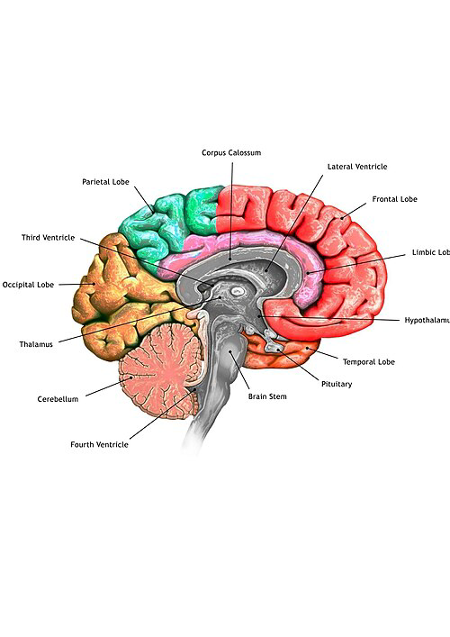
The brainstem can be divided into three sections: the midbrain (mesencephalon), pons (metencephalon), and medulla oblongata (myelencephalon). The medulla oblongata and pons, along with the cerebellum, are considered parts of the hindbrain (rhombencephalon); however, the cerebellum itself is not part of the brainstem.
Medulla Oblongata
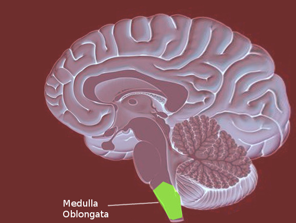
The Medulla Oblongata contains the nuclei (source) for the Glossopharyngeal (IX), Vagus (X) Hypoglossal (XII) and the Accessory nerves (Hurley et al., 2010). It also contains parts of the Trigeminal Nerve (V). Below the Medulla Oblongata lies the median fissure, connecting the brain to the spinal cord (Iordanova & Reddivari, 2023).
Functionally, the medulla regulates automatic responses such as sneezing, balance coordination, cardiovascular and respiratory systems, and other vital involuntary actions.
Pons
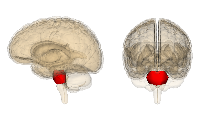
The pons, meaning "bridge" in Latin, is the middle section of the brainstem, positioned between the midbrain superiorly and the medulla oblongata inferiorly. It has an approximate height of 27 mm, transverse width of 38 mm, and anteroposterior width of 25 mm.
The pons contains the nuclei for the Trigeminal (V), Abducens (VI), Facial (VII), and Vestibulocochlear (VIII) cranial nerves. It acts as a relay station, transmitting signals from the cerebrum to the cerebellum via the pontocerebellar fibers, and coordinates essential functions such as respiration, sleep-wake cycles, facial expressions, hearing, and balance (Rahman & Tadi, 2020).
Midbrain
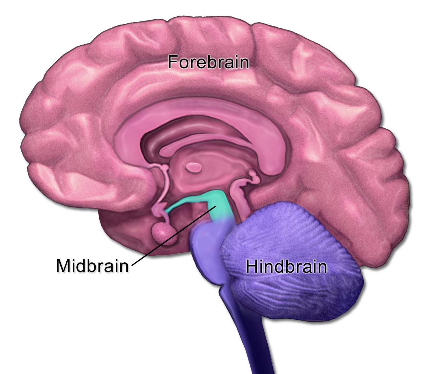
The midbrain, or mesencephalon, is the smallest portion of the brainstem at approximately 1.5 cm in length. It serves as the origin for the Oculomotor (III) and Trochlear (IV) cranial nerves, responsible for eye movements, eyelid elevation, and pupil constriction via the parasympathetic Edinger-Westphal nucleus. Although the Trigeminal nerve (V) primarily originates from the pons, its nuclei extend into the midbrain to mediate proprioceptive functions.
Anatomically, the midbrain is divided into the ventral tegmentum—containing the substantia nigra (dopamine production, motor control) and red nucleus (limb coordination)—and the dorsal tectum, which includes the superior and inferior colliculi for visual and auditory reflexes, respectively.
Functionally, the midbrain is critical for sensory-motor integration, consciousness through the reticular activating system, pain modulation via the periaqueductal gray matter, and reward pathways in the ventral tegmental area (Caminero & Cascella, 2024).
Cerebellum
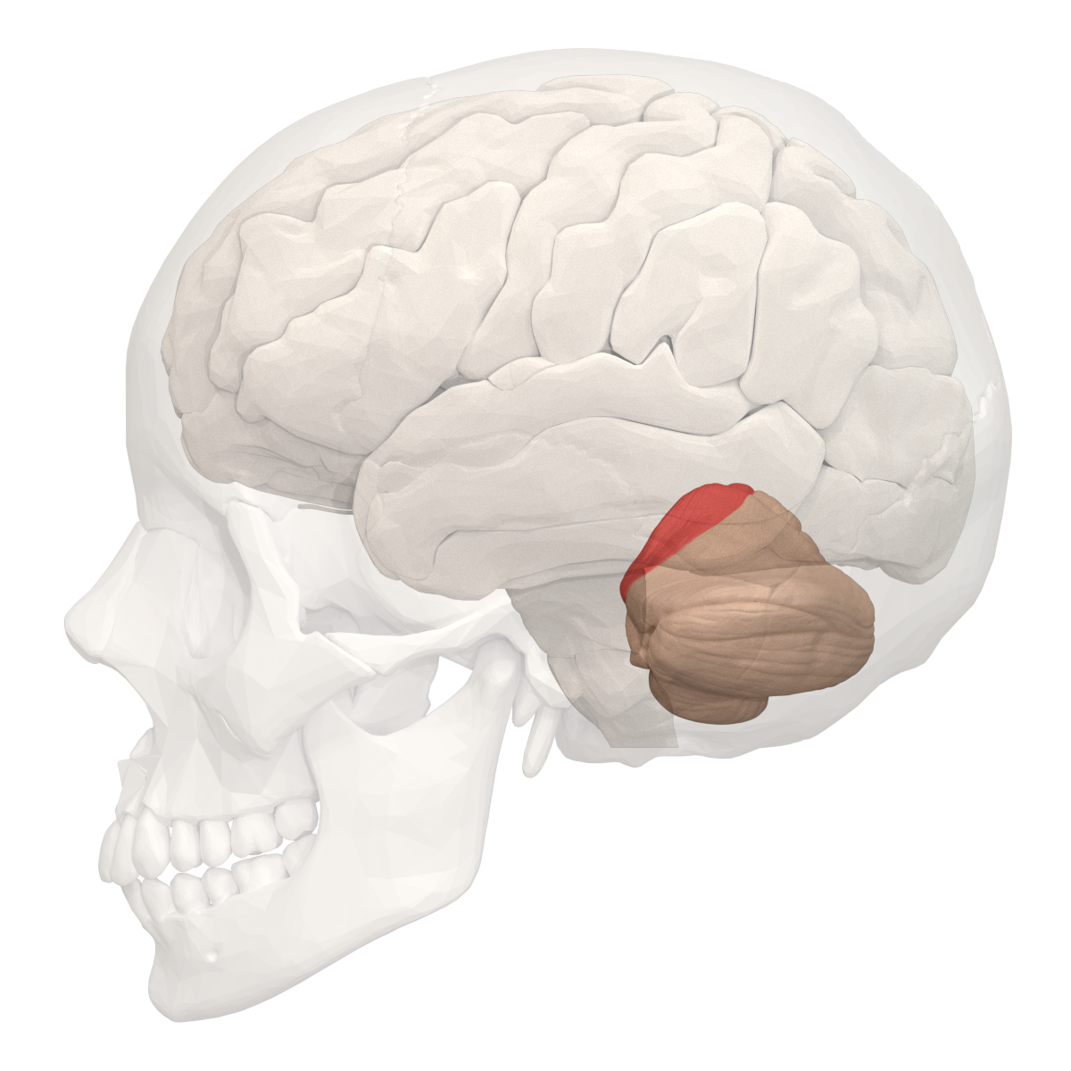
Although not part of the brainstem proper, the cerebellum is closely integrated with it as a key component of the hindbrain, located in the posterior cranial fossa behind the pons and medulla oblongata, separated from the cerebrum by the tentorium cerebelli. This structure, often called the "largest part of the hindbrain," consists of two hemispheres connected by the vermis and is divided into three lobes: anterior, posterior (the largest), and flocculonodular, separated by the primary and posterolateral fissures. It attaches to the brainstem via three pairs of cerebellar peduncles—superior, middle, and inferior—which facilitate bidirectional communication for coordinating motor activities. The cerebellum's primary functions include regulating motor movements, maintaining balance and posture, controlling muscle tone, and supporting motor learning, though it does not initiate voluntary actions. Deep within its white matter (the arbor vitae) lie three pairs of nuclei: fastigial (medial, for trunk coordination), interposed (globose and emboliform, for limb control), and dentate (lateral, for planning complex movements). Damage here can lead to ataxia, intention tremor, dysmetria, and other ipsilateral coordination deficits, underscoring its vital role in precise, adaptive movement (Jimsheleishvili & Dididze, 2023).
The "reptile brain" refers to the brainstem as the most ancient, evolutionarily conserved core of the vertebrate brain—shared with reptiles and responsible for primal, survival-driven instincts like the "fight or flight" response, heartbeat regulation, and reflexive behaviors.
As Carl Sagan implies, our passion for learning—harnessed by the neocortex—empowers us to modulate these instinctive drives, transforming raw reactivity into deliberate healing and adaptation in therapy and beyond.
References
- Caminero, F., & Cascella, M. (2024). Neuroanatomy, mesencephalon midbrain. In StatPearls [Internet]. StatPearls Publishing.
- Hurley, R. A., Flashman, L. A., Chow, T. W., & Taber, K. H. (2010). The brainstem: anatomy, assessment, and clinical syndromes. The Journal of neuropsychiatry and clinical neurosciences, 22(1), iv-7.
- Jimsheleishvili, S., & Dididze, M. (2023). Neuroanatomy, cerebellum. In StatPearls [Internet]. StatPearls Publishing.
- Iordanova R, Reddivari AKR. Neuroanatomy, Medulla Oblongata. In: StatPearls. StatPearls Publishing, Treasure Island (FL); 2025. PMID: 31869070.
- Parvizi, J., & Damasio, A. (2001). Consciousness and the brainstem. Cognition, 79(1-2), 135-160.
- Rahman M, Tadi P. Neuroanatomy (2020), Pons. In: StatPearls. StatPearls Publishing, Treasure Island (FL); 2025. PMID: 32809424.
- Venkatraman, A., Edlow, B. L., & Immordino-Yang, M. H. (2017). The brainstem in emotion: a review. Frontiers in neuroanatomy, 11, 15.