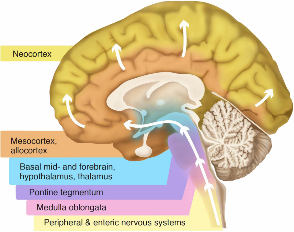
The cerebral cortex represents one of the most recently evolved structures of the brain, often referred to as the neo-mammalian brain. Understanding brain evolution involves comparing mammalian neuroanatomy, integrating evidence from the fossil record, and forming testable evolutionary theories. (Kaas, 2006).
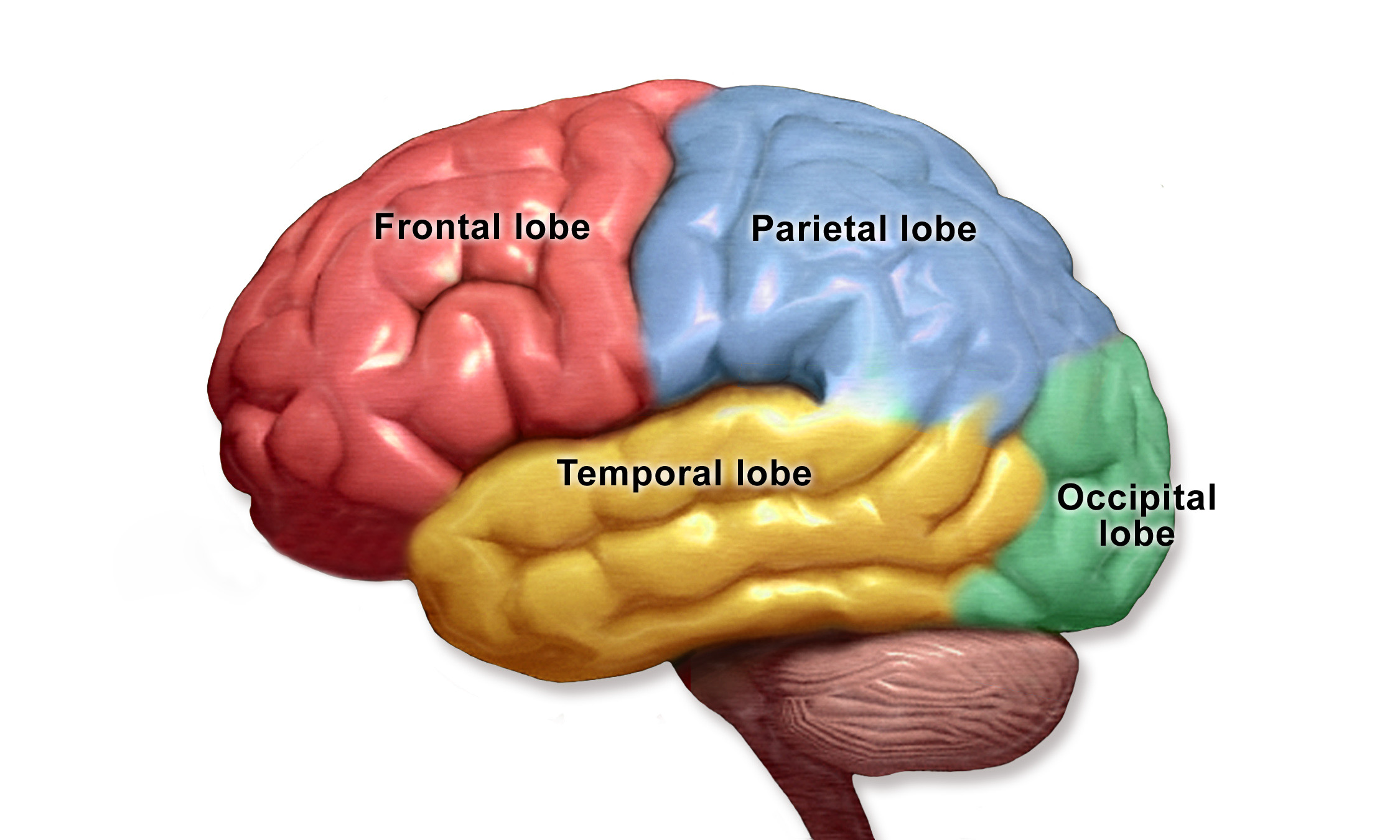
The human brain contains approximately 86 billion neurons and about ten times as many glial cells. Of these, the cerebral cortex contains around 16 billion neurons but accounts for about 80% of the brain's total mass (Azevedo et al., 2009). The cerebral cortex can be categorized into four main lobes, which are divided by key fissures and sulci (grooves), and differ in cell number, density, and morphology (Vacha et al., 2022; Cadwell et al., 2019).
Developmentally and anatomically, the cortex can also be divided into the neocortex (or isocortex)—which makes up about 90% of the cerebral cortex and supports higher-order cognitive functions such as language, reasoning, and consciousness—and the allocortex, the evolutionarily older part that includes structures of the limbic system (Cadwell et al., 2019).
Brain Lobes
Each of the four major lobes of the cerebral cortex plays a specialized yet interconnected role in human cognition and perception.
Temporal Lobe
Different systems of the brain evolved at varying rates and times in response to environmental pressures, a pattern known as mosaic evolution (Bruner et al., 2023; Schoenemann, 2012). The temporal lobe underwent significant evolutionary changes in early humans, coinciding with the emergence of social complexity and settled communities. This region is strongly associated with language, comprehension, and auditory processing.
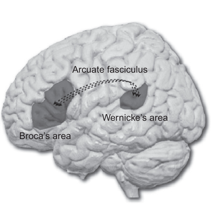
Wernicke's area, one of the first brain regions identified as being related to language, is primarily responsible for speech comprehension. In contrast, Broca's area, located in the inferior frontal gyrus of the frontal lobe, is involved in speech production. These two regions communicate via the arcuate fasciculus, a bundle of white matter fibers that enables the coordination necessary for complex verbal communication.
Beyond language, the temporal lobe also contributes to emotion, visual recognition, and memory. This is due to its close association with components of the limbic system, including the amygdala and hippocampus, which are situated within the temporal regions (Torrico & Abdijadid, 2023).
Occipital Lobe
The occipital lobe is the smallest of the four cerebral lobes and is located at the posterior region of the brain, just above the cerebellum. Visual information from the eyes is transmitted via the optic nerves, which partially cross at the optic chiasm—an X-shaped structure that connects the optic nerves to the optic tracts. This crossing allows visual input from each eye to be processed by the opposite hemisphere of the brain, resulting in coordinated left and right visual fields (Kidd, 2014).
As its name implies, the occipital lobe is primarily responsible for visual processing. It contains the primary visual cortex, which interprets visual features such as color, motion, shape, objects, and faces (Rehman & Khalili, 2023). Through its connections with other cortical regions, it also contributes to visual memory formation and recall.
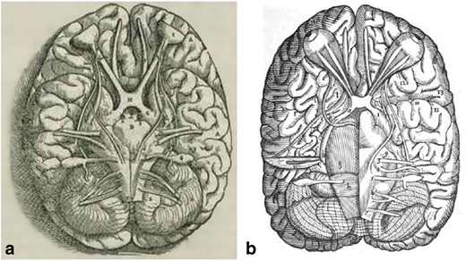
Source: Costea et al., 2017
Parietal Lobe
The parietal lobe acts as an integrative hub for sensory and cognitive processes. It receives and interprets information related to touch, pressure, pain, temperature, and proprioception, allowing the brain to construct an awareness of the body's position in three-dimensional space and navigate the surrounding environment. The primary somatosensory cortex, located in the postcentral gyrus, processes tactile and proprioceptive information, while the posterior parietal cortex integrates these inputs to guide spatial awareness and attention.
Beyond sensory integration, the parietal lobe also plays a crucial role in higher cognitive functions, including working memory, numerical reasoning, and mathematical cognition. The intraparietal sulcus (IPS), in particular, is strongly implicated in quantitative processing, mental calculation, and the manipulation of information in working memory (Dehaene et al., 2003; Koenigs et al., 2009; Whitlock, 2017). Through these combined sensory and cognitive functions, the parietal lobe helps coordinate perception, action, and abstract thought.
Frontal Lobe
The frontal lobe is located at the anterior region of the brain, as its name suggests. It is one of the last areas of the neocortex to fully develop and has undergone significant expansion during human evolution (Fuster, 2002). The frontal lobe is responsible for a range of complex cognitive and behavioral functions, including problem-solving, attention, working memory, language, decision-making, creativity, emotional regulation, impulse control, and self-awareness (Scott & Schoenberg, 2011).
Notably, regions within the medial prefrontal cortex are strongly associated with self-referential processing and the theory of self, which involves reflecting on one's own thoughts, emotions, and identity, as well as understanding oneself in relation to others (Northoff et al., 2006).
Comparative Cortical Development
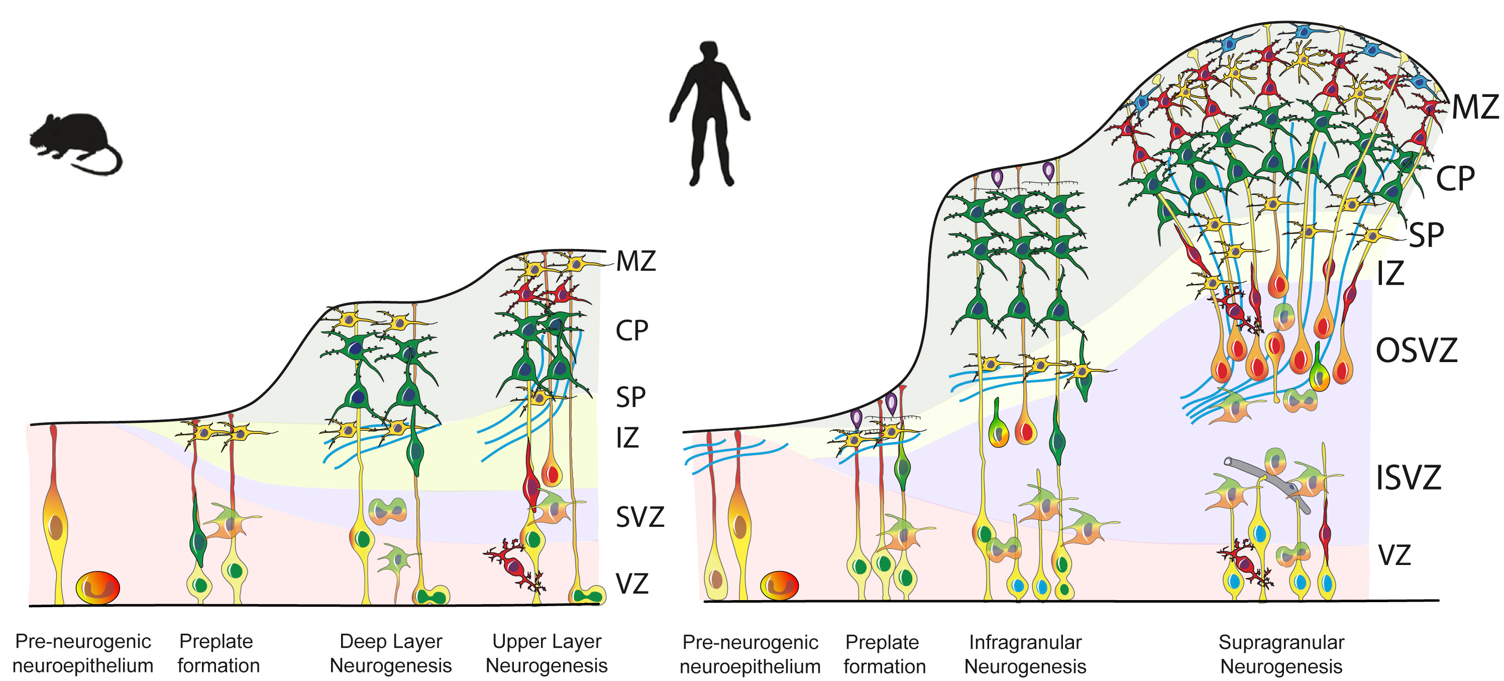
The illustration above highlights key differences between mouse and human cortical development. While both species share the same fundamental stages of neurogenesis, the human cortex exhibits an expanded outer zone and a greater diversity of progenitor cells. This expansion supports the increased number of cortical layers and the enhanced complexity of neural connectivity observed in humans. These structural differences reflect the evolutionary enlargement and specialization of the neocortex, particularly in regions such as the prefrontal cortex, which underlie advanced cognitive abilities including language, planning, and self-awareness.
Together, these developmental and structural adaptations underscore how evolution shaped the human brain into a system capable of integrating sensory, emotional, and abstract thought — the foundation of what we perceive as consciousness and the self.
Cerebral Cortex Lobes Overview
References
- Bruner, E., Battaglia-Mayer, A., & Caminiti, R. (2023). The parietal lobe evolution and the emergence of material culture in the human genus. Brain Structure and Function, 228(1), 145-167.
- Cadwell, C. R., Bhaduri, A., Mostajo-Radji, M. A., Keefe, M. G., & Nowakowski, T. J. (2019). Development and arealization of the cerebral cortex. Neuron, 103(6), 980-1004.
- Costea, C. F., Turliuc, Ş., Buzdugă, C., Cucu, A. I., Dumitrescu, G. F., Sava, A., & Turliuc, M. D. (2017). The history of optic chiasm from antiquity to the twentieth century. Child's Nervous System, 33(11), 1889-1898.
- Dehaene, S., Piazza, M., Pinel, P., & Cohen, L. (2005). Three parietal circuits for number processing. In The handbook of mathematical cognition (pp. 433-453). Psychology Press.
- Frederico A.C. Azevedo; Ludmila R.B. Carvalho; Lea T. Grinberg; José Marcelo Farfel; Renata E.L. Ferretti; Renata E.P. Leite; Wilson Jacob Filho; Roberto Lent; Suzana Herculano-Houzel. (2009). Equal numbers of neuronal and nonneuronal cells make the human brain an isometrically scaled-up primate brain. , 513(5), 532–541. doi:10.1002/cne.21974
- Fuster, J. M. (2002). Frontal lobe and cognitive development. Journal of neurocytology, 31(3), 373-385.
- Kaas, J. H. (2006). Evolution of the neocortex. Current Biology, 16(21), R910-R914.
- Kidd, D. (2014). The optic chiasm. Clinical Anatomy, 27(8), 1149-1158.
- Northoff, G., Heinzel, A., de Greck, M., Bermpohl, F., Dobrowolny, H., & Panksepp, J. (2006). Self-referential processing in our brain—a meta-analysis of imaging studies on the self. NeuroImage, 31(1), 440–457. https://doi.org/10.1016/j.neuroimage.2005.12.002
- Rehman A, Al Khalili Y. Neuroanatomy, Occipital Lobe. In: StatPearls. StatPearls Publishing, Treasure Island (FL); 2025. PMID: 31335040.
- Schoenemann, P. Thomas. (2012). [Progress in Brain Research] Evolution of the Primate Brain Volume 195 || Evolution of brain and language. , (), 443–459. doi:10.1016/b978-0-444-53860-4.00022-2
- Scott, J. G., & Schoenberg, M. R. (2010). Frontal lobe/executive functioning. In The little black book of neuropsychology: A syndrome-based approach (pp. 219-248). Boston, MA: Springer US.
- Torrico, T. J., & Abdijadid, S. (2023). Neuroanatomy, limbic system. In StatPearls [Internet]. StatPearls Publishing.
- Vachha, B. A., Massoud, T. F., & Huang, S. Y. (2022). Anatomy of the cerebral cortex, lobes, and cerebellum. Neuroimaging Clinics, 32(3), 463-473.
- Whitlock, J. R. (2017). Posterior parietal cortex. Current biology, 27(14), R691-R695.