Perception - Sight
Perception involves the constant accumulation of sensory inputs from body sensors, transmitted via nerves to the brain for processing and interpretation. Senses are absorbed through our body sensors but not only sensors come into play. Beyond the major senses of touch, vision, hearing and taste; our bodies can distinguish distances, momentum, balance and many other interoceptive (which relay internal messages, such as hunger) stimuli (Kassab & Alexandre, 2015). In this section of the site, we will initially present the basic functionality of our interoceptive and exteroceptive sensations, before diving into how all these sensations lead to the understanding of perception.
Let's first explore vision, which begins with light:
Eye Anatomy
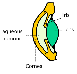
Cornea: The transparent, dome-shaped outer layer at the front of the eye covering the iris and pupil. It acts as the primary focusing element, refracting incoming light to form a clear image on the retina, maintaining hydration while protecting against dirt, germs, and UV light. Some techniques used to analyze and observe a living cornea includes In vivo confocal microscopy (IVCM) and Computerized videokeratography.
Aqueous Humor: The clear fluid in the front chamber (between cornea and lens), supplying nutrients, removing waste, and maintaining intraocular pressure. Surrounds the iris and lens of the eye.
Iris: The colored muscular ring surrounding the pupil. Rich in blood supply. It adjusts pupil size to regulate light entry, contracting in bright light (smaller pupil, Miosis) and relaxing in dim light (larger pupil, Mydriasis), functioning like a camera's aperture. This is possible due to loosely structured microfibers that allow fluid to enter and exit.
Lens: A flexible, transparent structure located behind the iris and pupil, responsible for refining the focus of light onto the retina to produce clear images. It dynamically adjusts its shape through contractions of the muscles, enabling accommodation for sharp vision at varying distances which plays a key role in depth perception. Composed primarily of tightly packed lens fibers, the lens is enveloped by the capsule, a thin, elastic membrane that provides structural support and anchors it to the ciliary muscles via zonules, allowing for precise shape changes. Beneath the capsule lies the epithelium, a single layer of cells that handles secretion and absorption, regulating nutrient uptake and waste removal while producing new lens fibers throughout life to maintain transparency and support growth and repair.
Sclera: The tough, white outer layer ("white of the eye") providing structural support and protection, covering the eye except the cornea.
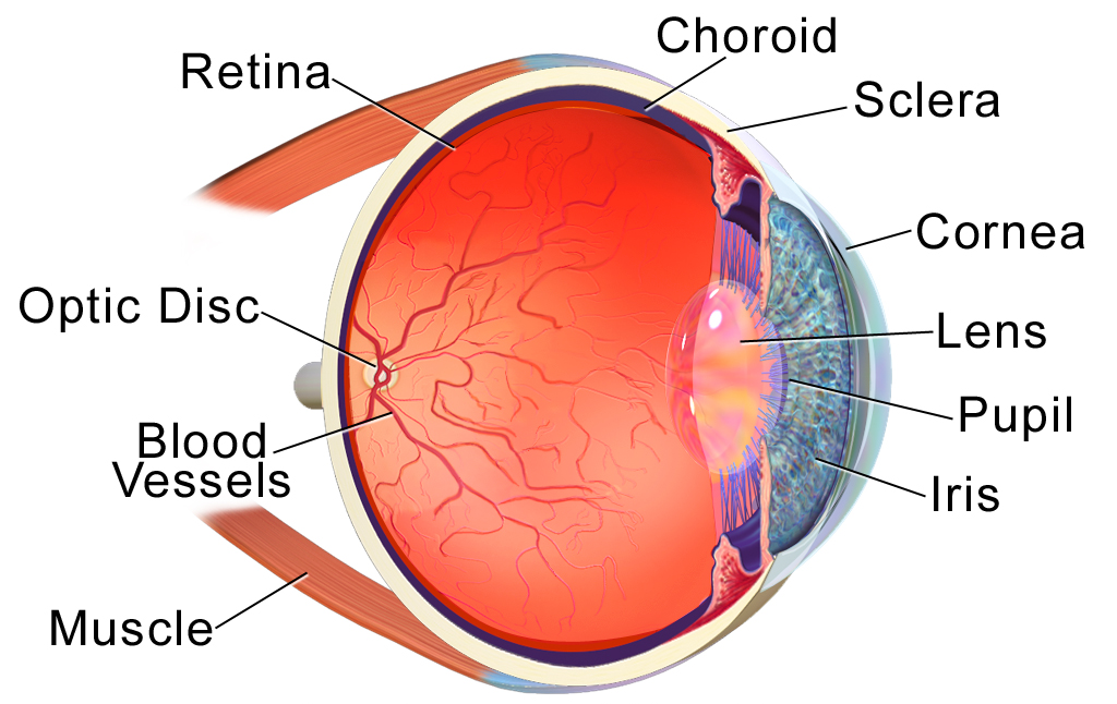
Pupil: The black circular opening in the iris center that permits light entry. Its size, controlled by the iris, optimizes light intake for clear vision.
Choroid: The vascular middle layer between the sclera and retina, supplying nutrients and oxygen via blood vessels and absorbing excess light to prevent internal reflections, preventing blurriness and glares.
Retina: The light-sensitive inner most layer at the eye's back with photoreceptors (rods for low-light/peripheral vision, cones for color/detail), converting light into electrical signals for brain interpretation.
Optic Disk: The point where the optic nerve exits the eye, carrying visual signals to the brain. It lacks photoreceptors, creating a natural blind spot in the visual field.
Macula Lutea: A small, central area of the retina rich in cones, responsible for sharp, detailed central vision and color perception, critical for tasks like reading or recognizing faces.
Optic Nerve: A bundle of nerve fibers transmitting electrical impulses from the retina to the brain's visual cortex. Damage, as in glaucoma, can cause vision loss.
Vitreous Humor: The clear, gel-like substance between the lens and retina, maintaining eye shape and aiding light transmission.
Light enters via the cornea, which refracts it, then passes through the pupil (regulated by the iris). The lens further focuses light onto the retina, particularly the macula lutea for sharp central vision. The retina converts light into electrical signals, sent via the optic nerve (exiting at the optic disk) to the brain for visual processing. This involves refraction, accommodation, and neural transmission for clear, detailed vision (Kassab and Alexandre, 2015).
Phototransduction: Converting Light to Neural Signals
The process of which light energy is transformed into electrical signals is called Phototransduction and occurs in the retina. It allows us to perceive the visual world. Specialized cells like rods, which excel in low-light vision, and cones, which handle color and detail, contain photopigments that absorb photons. This triggers a biochemical cascade: the pigment molecule changes shape, closing ion channels and hyperpolarizing the cell to generate an electrical impulse. The signal is then relayed through bipolar cells, which process and refine the information, before passing it to ganglion cells. Finally, the ganglion cells bundle their axons into the optic nerve, transmitting the signals to the brain for interpretation, enabling everything from detecting motion to recognizing shapes. Fun fact, the average human reaction time is around 250 milliseconds (Mazumdar et al., 2014).
Recognizing the Color Spectrum
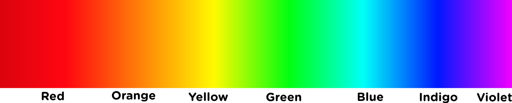
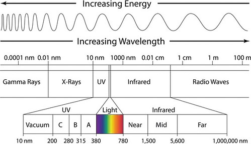
The eye distinguishes the full color spectrum—ranging from red (longer wavelengths around 700 nm) to violet (shorter wavelengths around 400 nm), as shown in the visible light gradient—through three types of cone photoreceptors in the retina, each sensitive to different wavelengths. Short-wavelength cones (S-cones) detect blues and violets, medium-wavelength cones (M-cones) respond to greens and yellows, and long-wavelength cones (L-cones) pick up reds and oranges. When light from the spectrum hits the retina, these cones activate in varying combinations based on the light's wavelength; for instance, yellow light stimulates both M- and L-cones equally, while indigo excites S- and M-cones more. The brain interprets these overlapping signals via trichromatic color vision theory, blending them to perceive millions of hues, though illusions or deficiencies (like color blindness) can alter this perception. This system ensures we see the vibrant rainbow of colors in everyday life.
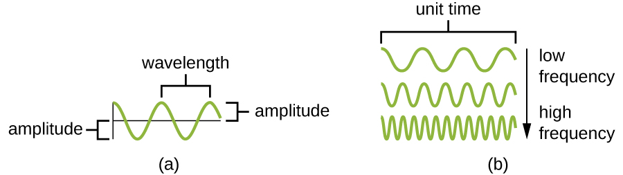
The electromagnetic spectrum encompasses a wide range of light wavelengths and frequencies, from long radio waves to short gamma rays, with visible light—a tiny speck—detected by our eyes through phototransduction. As illustrated, wavelength (the distance between wave peaks, as shown in the diagram above (a)) and frequency (the number of waves per unit time, (b)) define this spectrum, where shorter wavelengths (e.g., violet at ~400 nm) correspond to higher frequencies, and longer wavelengths (e.g., red at ~700 nm) to lower frequencies (Zwinkels, 2023). In the retina, photoreceptors like rods (for low-light vision) and cones (for color) absorb these wavelengths, triggering a cascade where photopigments like rhodopsin or photopsins change shape, close ion channels, and generate electrical signals. This signal, relayed through bipolar and ganglion cells to the optic nerve, allows the brain to interpret the vibrant color spectrum—reds, greens, blues, and all hues in between—based on the combined activation of cone types sensitive to different frequencies, shaping our perception of the world.
Brightness and Amplitude in Visual Perception
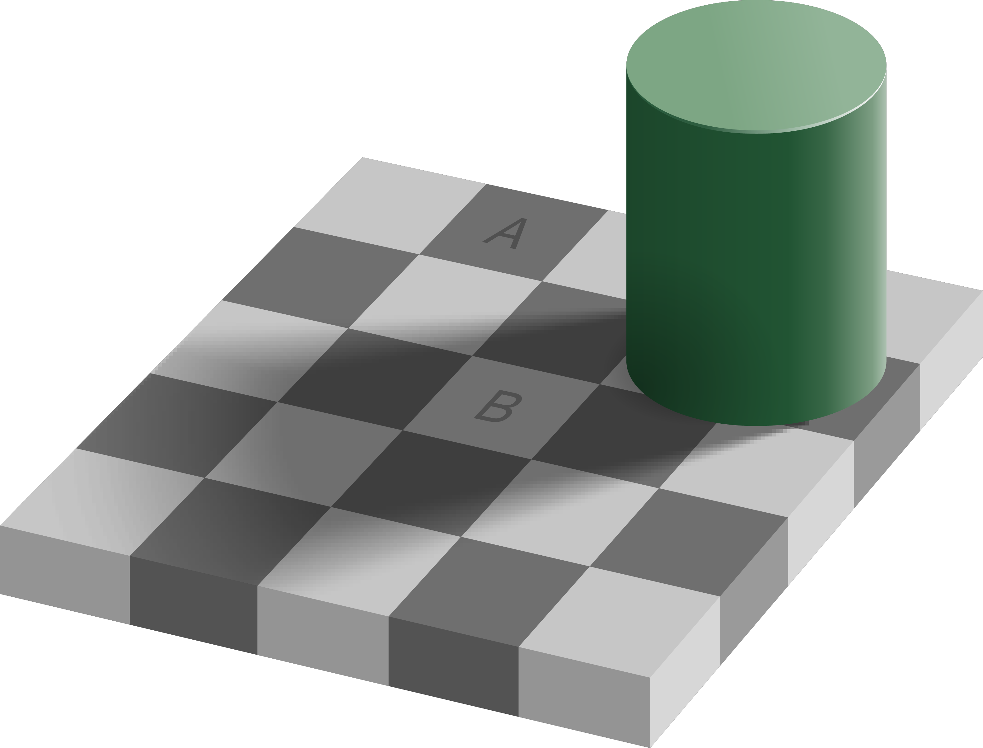
Brightness, or the perceived intensity of light, is directly influenced by the amplitude of light waves in the electromagnetic spectrum. Higher amplitude means greater energy in the wave, delivering more photons to the retina, which rods and cones interpret as brighter light; for example, a high-amplitude red wave appears as a vivid scarlet, while low amplitude dims it to a faint glow. This ties into phototransduction, where stronger signals from photoreceptors amplify the electrical impulse, allowing the brain to distinguish subtle shades in low light (via rods) or vibrant details in bright conditions (via cones). Interestingly, our perception of brightness isn't linear—context like surrounding darkness can create illusions. These brightness illusions are exemplified in phenomena like the Checker Shadow Illusion.
Fun Optical Illusions and How They Work
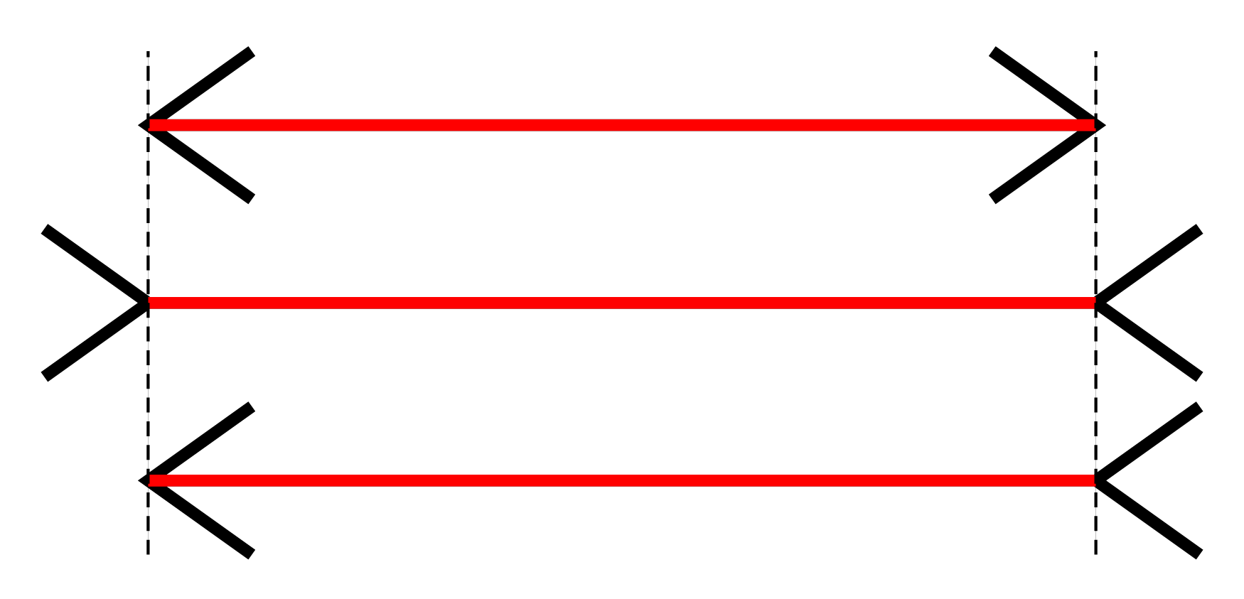
Müller-Lyer Illusion
The brain uses depth cues (like arrows suggesting corners) to overestimate or underestimate length, based on perspective interpretation.
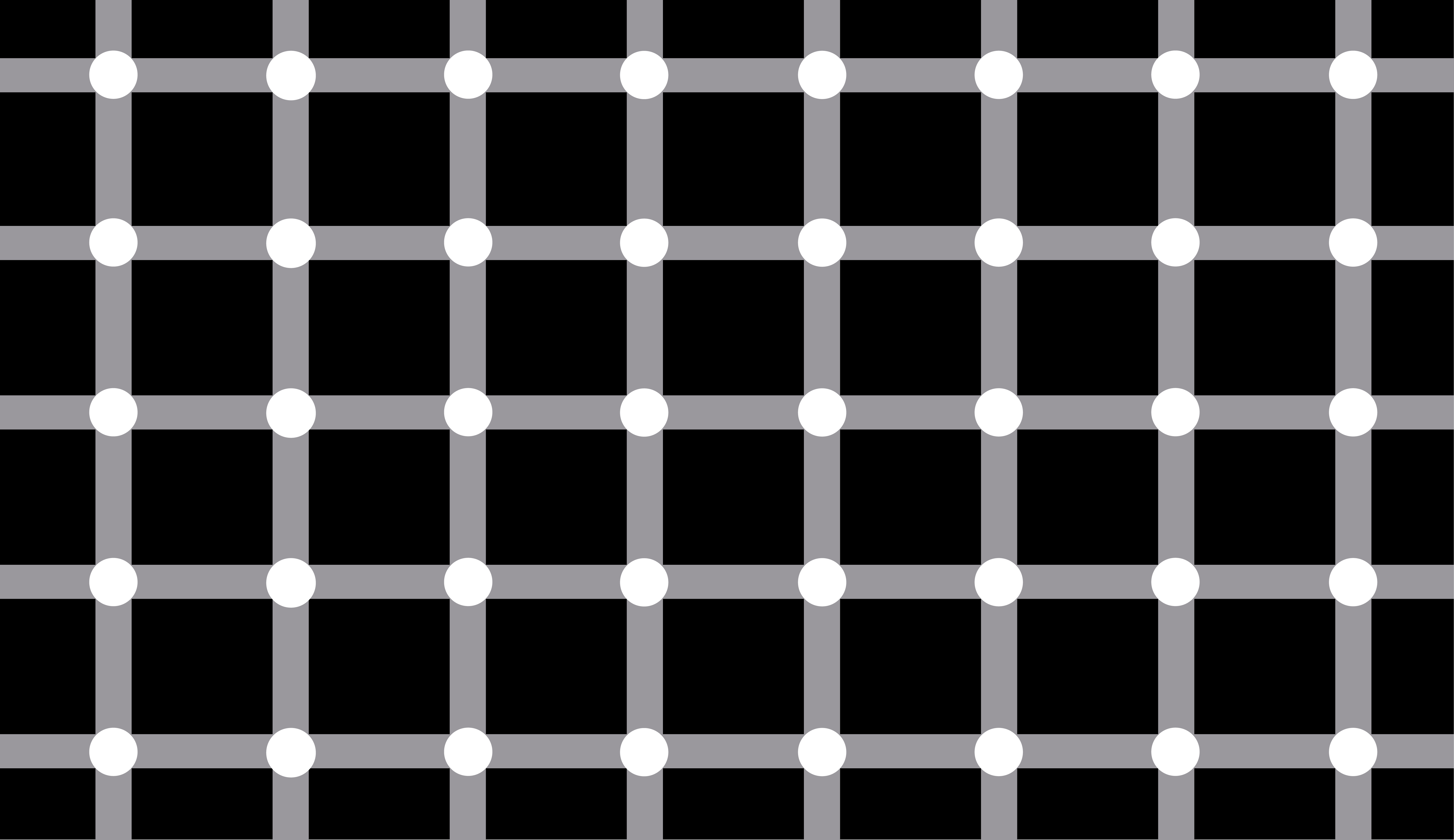
Scintillating Grid
Lateral inhibition in retinal cells enhances contrast at edges, causing "ghost" dots from overstimulation of peripheral vision.
Checker Shadow Illusion
The brain adjusts for lighting context (shadows), perceiving the shaded square as lighter to maintain color constancy.
Mirages
Atmospheric refraction bends light rays through layers of air with varying temperatures and densities, creating false reflections of the sky or distant scenes on the horizon, tricking the eye into seeing displaced or mirrored images.

References
- Forrester, J. V., Dick, A. D., McMenamin, P. G., Roberts, F., & Pearlman, E. (2021). The eye. Elsevier.
- Kassab, R., & Alexandre, F. (2015). Integration of exteroceptive and interoceptive information within the hippocampus: a computational study. Frontiers in Systems Neuroscience, 9, 87.
- Mazumdar, D., Pel, J. M., Panday, M., Asokan, R., Vijaya, L., Shantha, B., ... & Van Der Steen, J. (2014). Comparison of saccadic reaction time between normal and glaucoma using an eye movement perimeter. Indian journal of ophthalmology, 62(1), 55-59.
- Zwinkels, J. (2023). Light, electromagnetic spectrum. In Encyclopedia of color science and technology (pp. 1062-1069). Cham: Springer International Publishing.
Images for this article were collected from Wikimedia Commons and Flickr. Images were slightly edited for use.